
Digital X-ray in Eluru
A digital X-ray is an advanced imaging technology that captures internal body structures using electronic sensors instead of traditional photographic film. Digital X-ray in Eluru offers rapid image acquisition, enhanced image quality, and easier storage and sharing of results through digital formats. Digital X-rays reduce radiation exposure for patients and allow for immediate review and diagnosis by healthcare providers. Their versatility makes them essential in modern medical diagnostics, providing detailed insights into bones, tissues, and organs with greater efficiency and precision compared to conventional X-ray methods.
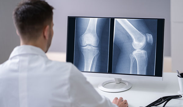
Advantages of Digital X-ray in Orthopedics
Digital X-ray machines significantly improve orthopedic imaging by providing rapid, high-resolution images that enhance diagnostic accuracy and treatment planning, especially in emergency cases like fractures. Their ability to instantly produce images reduces patient wait times and allows for immediate assessment, while image manipulation tools like zoom and contrast enhance clarity without additional scans. Digital systems also promote seamless electronic storage and sharing, fostering better collaboration among healthcare providers and streamlining workflows. They operate at lower radiation doses, making them safer for patients, particularly for pediatrics or repeated imaging needs. Environmentally, digital X-rays eliminate chemical processing, reducing hazardous waste. Although initial costs are higher compared to traditional film-based systems, digital X-ray technology offers long-term savings through decreased material expenses and increased durability, reducing the need for frequent replacements. Overall, bone injury X-ray in Eluru revolutionize orthopedic diagnostics by offering safer, faster, and more precise imaging solutions that improve patient care, enhance clinical efficiency, and support environmentally sustainable practices. This modern approach ultimately leads to better treatment outcomes and more efficient healthcare delivery. By searching for digital X-ray test near me in Eluru in your search engine you can ensure prompt diagnosis at our state of the art facility.
The Process
A digital X-ray is taken by positioning the patient between an X-ray source and a digital detector, often using a handheld or stationary device, depending on the area being examined. Once the patient is correctly aligned, the radiologic technologist activates the X-ray machine, which emits a controlled burst of radiation that passes through the body. The digital detector captures the transmitted X-rays, converting them into electronic signals almost instantaneously. These signals are then processed by a computer to produce high-resolution digital images that can be viewed immediately on a screen. This method offers advantages such as reduced radiation exposure, faster image acquisition, and enhanced image quality with adjustable contrast and brightness. The digital images can be easily stored, shared, and enhanced for detailed analysis, making the process efficient, safer, and more precise than traditional film-based X-rays.
Discover expert orthopedic care with Dr. I C Chenchaiah at Amulya Hospital, where cutting-edge technology meets compassionate treatment. A renowned orthopedician in Tadepalligudem, Dr. Chenchaiah utilizes the latest digital X-ray machines at Amulya Hospital to diagnose and treat musculoskeletal issues with unmatched accuracy.